Figure 8.
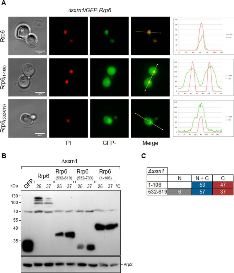
Deletion of SXM1 strongly affects the subcellular localization of Rrp6 mutants. A, fluorescence microscopy for the analysis of the subcellular localization of GFP-Rrp6 mutants in Δsxm1 cells growing at 25 °C. Mutants Rrp61–106 and Rrp6(532–619) were analyzed in this strain. Quantification is shown on the right. B, Western blot for the determination of the expression of the GFP-Rrp6 mutants in the Δsxm1 strain. Arp2 was used as an internal control. Mutants are expressed at higher levels than full-length Rrp6, and their levels do not decrease at 37 °C. C, quantitative analysis of Δsxm1 cells expressing GFP-fused Rrp6 mutants. N, protein concentrated in the nucleus; N + C, protein present both in nucleus and cytoplasm; C, protein visible mainly in the cytoplasm. Numbers correspond to the percentage of cells showing each phenotype. Scale bars, 4 μm.

