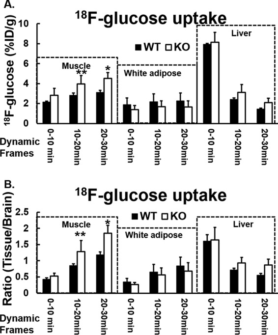
Figure 4.

In vivo insulin-stimulated [18F]FDG distribution in mice fed with normal chow diet is shown. A and B, after mice were fasted overnight, [18F]FDG 11.1 MBq (300 Ci) and insulin (1.125 units/kg) in 200 μl of saline were injected via tail vein under anesthesia. Dynamic imaging was acquired for 30 min by Siemens Inveon Small Animal PET/CT Imaging System. The data were obtained by drawing regions of interest (ROI) over target organs of muscle, abdominal subcutaneous white adipose tissue, liver, and brain (98, 99). The [18F]FDG bio-distribution in the target organs/tissues were analyzed at three time points: 10, 20, and 30 min. A, time-dependent distribution percentage of injected dose per gram (%ID/g) of [18F]FDG in the hind limb, liver, and abdominal white fat in Irak1 k/o mice and in wild-type mice. B, relative time-dependent distribution of [18F]FDG in the hind limb, liver, and abdominal white fat (ratio of [18F]FDG:specific tissue/brain) in Irak1 k/o mice and in wild-type mice. WT, n = 3; k/o, n = 3; *, p < 0.04; **, p < 0.03. Data shown are mean ± S.E.
