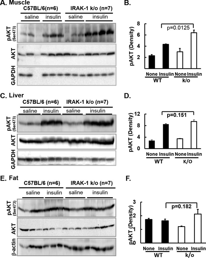
Figure 6.

Immunoblotting analysis of insulin signaling pathways in muscle, liver, and abdominal white adipose tissue is shown. A and B, muscle. C and D, liver. E and F, abdominal white adipose tissue. 20-week-old mice fed with chow diet were fasted for 5 h and then given saline (none) or insulin (10 units/kg body weight) via portal vein injection under anesthesia. 5 min after injection, skeletal muscle from hind limb, abdominal fat, and liver were immediately flash frozen and homogenized. These samples were centrifuged and supernatants of tissue lysates were analyzed by immunoblotting with indicated antibodies. A, C, and E, immunoblots with samples from 6 WT and 7 Irak1 k/o mice. B, D, and F, immunoblots for phospho-Akt were quantified by scanning densitometry and normalized for Akt total protein. Data are expressed as mean ± S.E.
