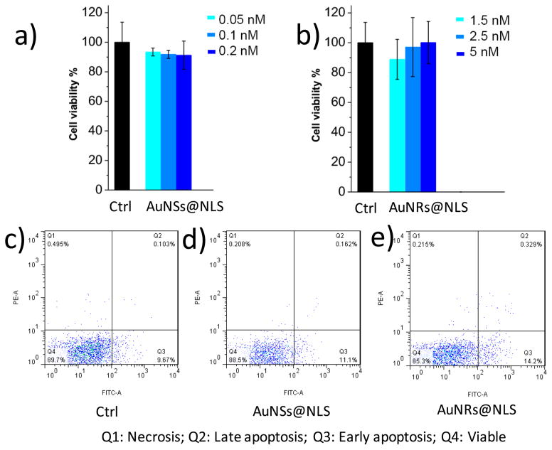
Figure 2.
Au nanoparticles cytotoxicity measurements and cellular uptake. (a) Cell viability measurement (XTT assay, n = 3) of HEY A8 cells after 24 h incubation with AuNSs@NLS at concentrations 0.05 nM (light blue), 0.1 nM (medium blue) and 0.2 nM (dark blue). (b) Cell viability (XTT, n = 3) assay for cells after 1.5 nM (light blue), 2.5 nM (medium blue) and 5 nM (dark blue) of AuNRs@NLS incubation with HEY-A8 cells for 24h. (c, d, and e) Flow cytometry experiment for apoptosis/necrosis assay (c, Ctrl; d, cells incubated with 0.2 nM of AuNSs@NLS; e, cells incubated with 5 nM of AuNRs@NLS).

