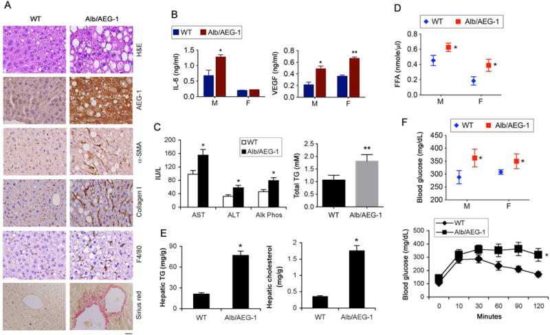
Figure 1.

Alb/AEG-1 mice develop NASH. A. H&E, IHC staining with the indicated antibodies and picrosirius red staining in FFPE sections of livers of 6 months old male littermates. Magnification 400×. Scale bar: 20 μm. B–E. IL-6 and VEGF (B), liver enzymes and total TG (C), and free fatty acid (FFA) (D) levels were measured in the sera and hepatic TG and cholesterol levels (E) were analyzed in 6-month old male littermates (n = 6/group). F. Blood glucose level was measured either under basal condition (top) or after glucose tolerance test (bottom). Male mice were used (n = 12/group). For graphs, data represent mean ± SEM. * : p<0.01, ** : p<0.001.
