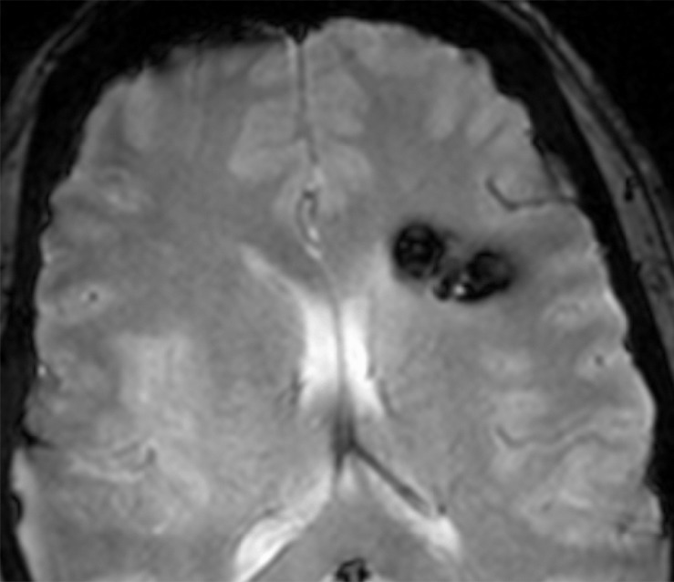
Figure 1a:

Images in 36-year-old woman with CCM1-CHM genotype who presented with symptomatic intracerebral hemorrhage. (a) Axial T2-weighted GRE MR image shows left frontal cavernous malformation. Patient had 10 additional brain lesions (not pictured). (b) Coronal reconstruction image from unenhanced abdominal CT performed for renal colic 3 years earlier reveals focal calcifications without associated mass in left adrenal gland.
