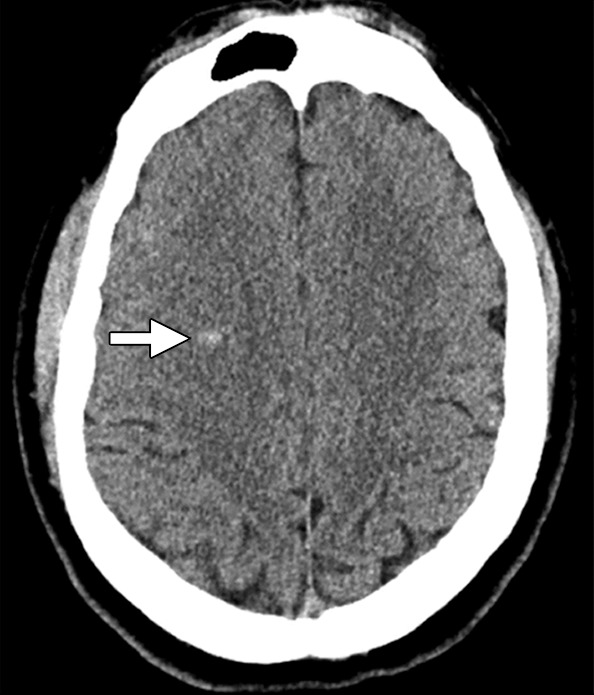
Figure 5a:

Images in 55-year-old man without prior neurologic symptoms who underwent imaging after fall from a horse. (a) Unenhanced axial head CT scan shows high-attenuation focus in right frontal lobe (arrow), which was interpreted as acute hemorrhage versus pre-existing cavernous malformation. (b) Contrast-enhanced axial CT scan of abdomen shows bilateral adrenal calcifications, which increased suspicion for fCCM. (c) Axial T2-weighted GRE brain MR image obtained at 1-month follow-up reveals multiple cavernous malformations, which confirms diagnosis of fCCM.
