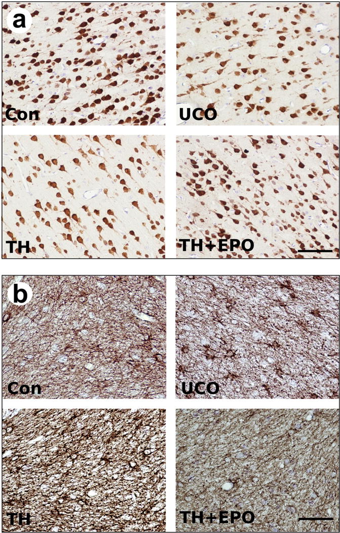
Figure 4.

Representative images of immunohistological staining for a) NeuN and b) GFAP in the cerebellum for control cesarean delivered animals (Con), asphyxiated animals (UCO), asphyxiated animals treated with therapeutic hypothermia (TH) and asphyxiated animals treated with therapeutic hypothermia plus Epo (TH+Epo). Scale bar = 70um.
