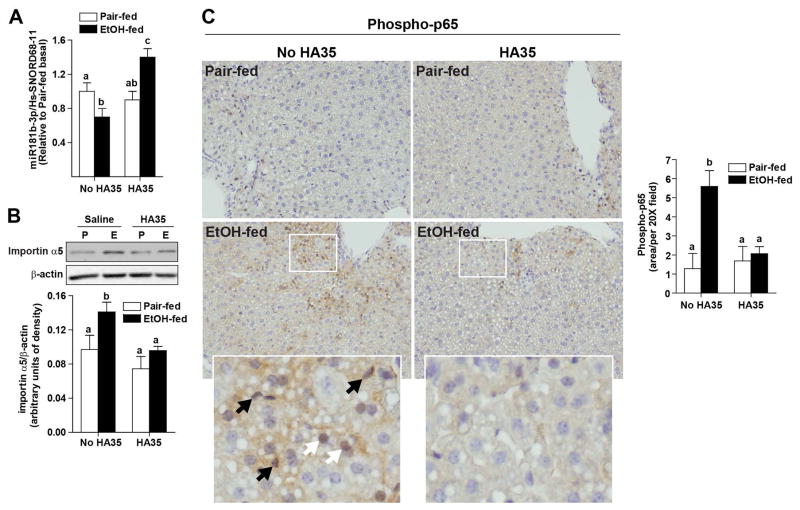
Figure 4. Regulation of importin α5 expression by ethanol and HA35 in an in vivo mouse model of ethanol-induced liver injury.
C57BL/6J mice were allowed free access to an ethanol containing diet for 4 days (2 days at 1% (v/v) ethanol, followed by 2 days 6% (v/v) ethanol) or pair-fed an isocaloric control diet. Mice were gavaged once daily for the last 3 days of ethanol feeding with HA35 at 1.5mg/kg body weight or saline control the last three days of the study. (A) Expression of miR181b-3p in whole liver was determined by qRT-PCR and normalized to Hs-SNORD68-11. (B) Non-parenchymal cells were isolated from livers by collagenase digestion and differential centrifugation. Cells were lysed and expression of importin α5 measured by Western blot and normalized to β-actin. (A–B) n=4 pair-fed and n=5–6 ethanol-fed. (C) Formalin-fixed paraffin-embedded sections of liver were de-paraffinized and localization of phospho-p65 was assessed by immunohistochemistry. Images were acquired at 20X. Black arrows on zoomed insets identify phospho-p65 in non-parenchymal cells while white arrows indicate hepatocytes. Images are representative of duplicate images from 4 pair-fed and 5–6 ethanol-fed mice in each treatment group.

