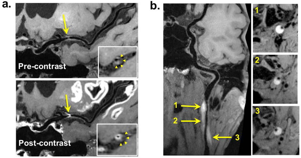
Figure 4.
Example patient images with curved multi-planar reconstruction. a) In a 58-year-old male patient, intracranial vessel wall (IVW) imaging reveals a focal lesion at the left middle cerebral artery M1 segment. It is characterized by eccentric wall thickening (arrows), suggestive of a plaque, on pre-contrast images and strong enhancement (arrows) at the plaque-lumen interface instead of in the entire thickened wall (arrowheads in the zoomed-in cross-sectional images) on post-contrast images. b) In a 51-year-old male patient, dissection at the left internal carotid artery, IVW imaging reveals a lesion (arrows) at the left internal carotid artery with hyper-intense, crescent-shaped intramural hematoma and longitudinally diffuse involvement.

