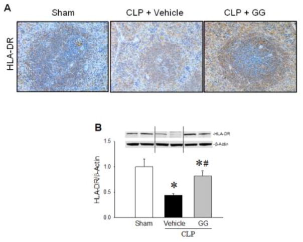
Figure 7.
Expression of HLA-DR in the spleens of septic aged rats. Aged rats subjected to sham operation or CLP with vehicle (normal saline) or GG (80 nmol/kg of Ghr and 50 μg/kg of GH) treatment at 5 h after CLP. Spleens were harvested at 20 h after CLP. Sections of splenic tissues were immunostained for HLA-DR. Positively-staining cells appear brown. Representative images of stained spleen sections are shown (A). In addition, HLA-DR levels in spleen tissues were measured by Western blotting (B). Data are expressed as mean ± SEM (n = 4–5 per group). * P < 0.05 versus sham and #P < 0.05 versus vehicle-treated septic animals.

