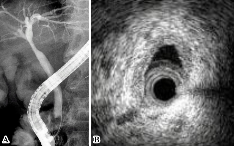
Figure 4.

(A) Endoscopic retrograde cholangiography revealed a slightly stenotic lesion of the hilar bile duct, and we conducted transpapillary bile duct brush cytology and a biopsy of the hilar bile duct stricture. (B) Transpapillary intraductal ultrasonography of the bile duct indicated a continuously thick wall from the upper to the lower bile duct with a smooth circular symmetric outer margin, a smooth inner margin, and a homogenous internal echo pattern.
