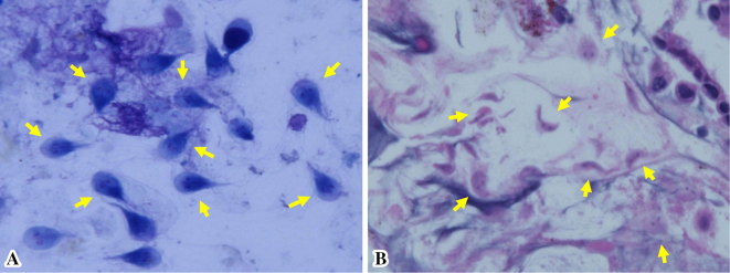
Figure 5.
A histopathological evaluation due to the findings on transpapillary bile duct brush cytology (A) and the biopsy (B). (A) Numerous active trophozoites of Giardia lamblia (arrows) (Giemsa staining×100). (B) Numerous active trophozoites of Giardia lamblia (arrows) in the bile duct wall but no malignant findings (Hematoxylin and Eosin staining ×100).

