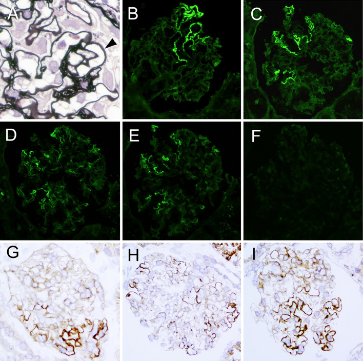
Figure 1.
Light micrograph of a part of the glomerulus, showing spikes (arrow head) of the basement membrane in the segmental capillary loops (A). Immunofluorescent findings show segmental positive granular IgG (B), IgG1 (C), κ light chain (D) and λ light chain (E) staining along the glomerular capillary walls and negative M-type phospholipase A2 receptor staining (F). Indirect immunohistochemistry for IgG shows granular staining along the segmental glomerular capillary walls with a variety of distribution (G-I). A: periodic acid-methenamin silver. Original magnification 600×, B-I: Original magnification 200×. G-I; Polyclonal anti-human IgG (DAKO Japan, Japan) was used.

