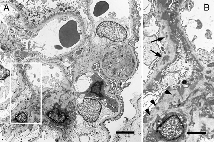
Figure 2.
Electron-microscopic findings. Normal capillary loop (right lower part) and capillary loops with subepithelial electron dense deposits (EDDs) can be seen (A). The area of the square in Fig. 2A shows not only subepithelial EDDs but also intramembranous EDDs (arrows) and mesangial EDDs (arrow heads). A; Bar=5 μm, B; Bar=2 μm.

