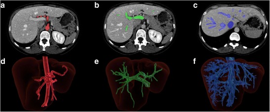
Fig. 15.

Liver subsegmentation according to vascular anatomy. Axial enhanced CT showing the segmented a hepatic arterial, b portal venous and c hepatic venous structures. Three-dimensional rendering of the same liver showing the corresponding segmented d arterial, e portal venous and f hepatic venous structures [13]
