
Fig. 4.
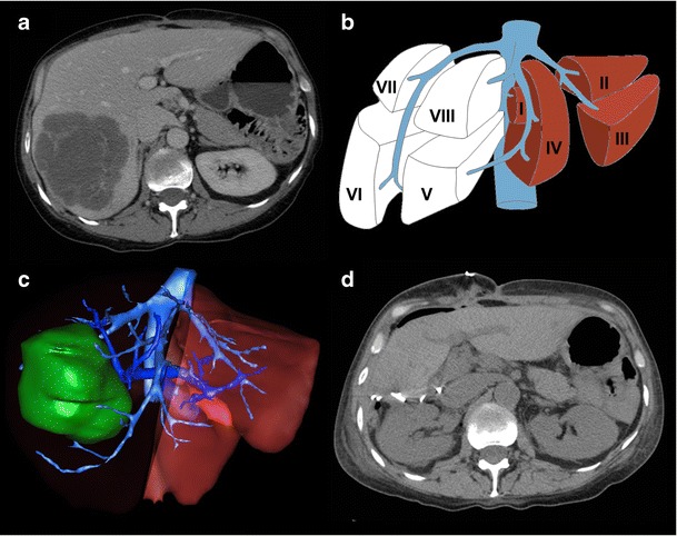
Future liver remnant volume calculation in normal liver prior to right hepatectomy. a Axial enhanced CT image showing colorectal liver metastasis involving right posterior segments (VI and VII). b Resection diagram shows the intended complete right hepatectomy surgery planned. c Three-dimensional rendered image showing surgical planning for complete right hepatectomy. FLR/TLV ratio was estimated to be 33%. d Axial unenhanced CT image of the same patient shortly after complete right hepatectomy. Actual FLR/TLV ratio was calculated to be 36%. Figure courtesy of Dr. Vandenbroucke-Menu; created with 3DVSP (IRCAD, Strasbourg, France)