
Fig. 5.
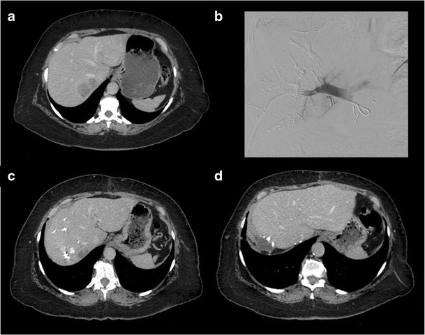
Portal vein embolisation prior to right hepatectomy. a Axial enhanced CT image shows colorectal liver metastasis involving segments V, VI, VII (only VII shown). b Final portogram of embolised portal vein branches in segments V through VIII using a Lipiodol-glue mixture. c Axial enhanced CT image obtained 1 month after right PVE showing hypertrophy of future liver remnant. d Axial enhanced CT image of the same patient after right hepatectomy