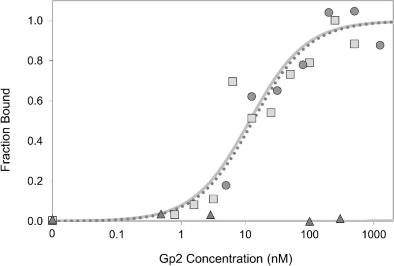
Fig. 2. Affinity titration.
A431 cells were labeled with DOTA-Gp2 domains (squares, DOTA-Gp2-EGFR; triangles, DOTA-Gp2-nb) at the indicated concentrations. Binding was detected by fluorescein-conjugated anti-His6 antibody via flow cytometry. Fluorescence signal is normalized between minimal and maximal fluorescence. One representative titration of triplicate experiments is presented. A representative Gp2-EGFR titration curve is also included for comparison (circles, dotted line). The equilibrium dissociation constant for DOTA-Gp2-EGFR, assuming a 1:1 binding model, is 7±5 nM

