Figure 3.

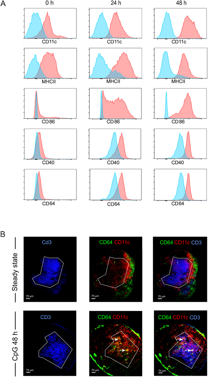
Phenotypic characterization and intranodal distribution of Ly6Chi monocytes. (A) Phenotypic characterization of Ly6Chi monocytes in the draining lymph node in response to subcutaneous CpG injection. The levels of CD11c, MHCII, CD86, CD40 and CD64 of ly6Chi monocytes are shown at 0 h, 24 h and 48 h after subcutaneous CpG injection. (B) Confocal images of popliteal lymph nodes in steady state and following footpad injection of CpG. Sections were stained for CD3 (blue), CD11c (red) and CD64 (green) to visualize T cells, DCs and Ly6Chi monocytes.
