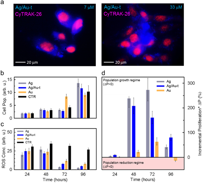
Figure 4.

(a) Confocal fluorescence microscope images of fixed NIH/3T3 fibroblast cells co-stained with Ag/Au-t clusters (blue) and CyTRAK-26 dye (red) under UV excitation at increasing cluster concentration (7 µM and 33 µM). (b) Cytotoxicity test and (c) ROS concentration level test on NIH/3T3 cells stained with 7 µM of Ag, Ag/Au-t and Au clusters, taken at four time-points during cells proliferation (24 h, 48 h, 72 h and 96 h). (d) Cellular proliferation test on cells in artificially stressed culture conditions by adding the metabolic accelerator menadione. The histogram shows the incremental cell proliferation of cells stained with the metal clusters calculated with respect to the unstained control culture.
