
Figure 3.
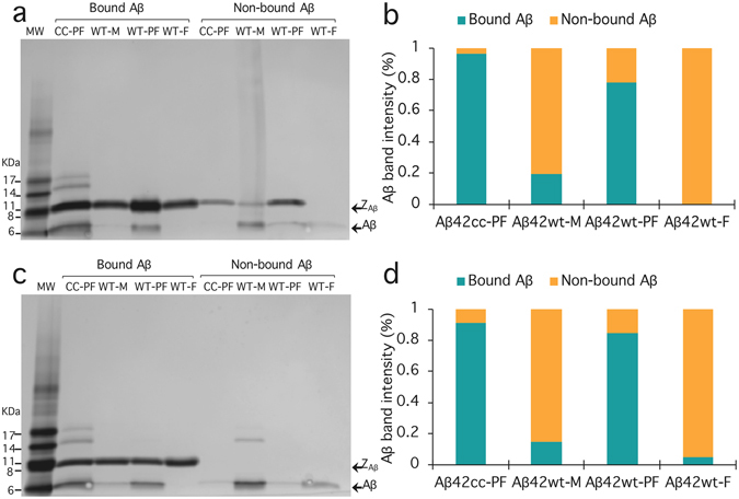
Binding of Affibody molecules to various Aβ aggregates and monomer in a batch mode affinity experiment. The SDS-PAGE analysis shows that Affibody ZA β 42cc_1 (a) and ZA β 42cc_4 (c) recognize protofibrillar aggregates of Aβ42cc and Aβ42wt (bound Aβ). Wild type monomer and fibrils did not bind to the Affibody molecules and were recovered in the supernatant (non-bound Aβ). The designations MW, CC-PF, WT-M, WT-PF and WT-F refer to molecular weight marker (GE Healthcare), Aβ42cc protofibrils, Aβ42wt monomer, Aβ42wt protofibrils and Aβ42wt fibrils, respectively. (b) and (d), Aβ band intensity for ZA β 42cc_1 and ZA β 42cc_4, respectively. The band intensities were obtained with the graphic program ImageJ32. The data was normalized with respect to each Aβ-species, i.e. the sum of the intensities for bands originating from one Aβ-species is equal to 100%.