
Figure 7.
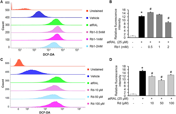
Rb1 or Rd treatment decreased atRAL-induced intracellular ROS production in ARPE19 cells. ARPE19 cells were treated with vehicle (saline or DMSO), Rb1 or Rd at the indicated concentrations for 1 h before being exposed to atRAL at 25 μM for 5 h, followed by DCF-DA staining for 30 min. After DCF-DA staining and washing, cells were resuspended and subject to further analysis by flow cytometry at the excitation and emission wavelengths of 490 nm and 520 nm, respectively. For each sample, 20,000 cells were acquired for data collection. Representative histograms of flow cytometry after DCF-DA staining were shown in (A,C). Relative DCF-DA fluorescence intensity was calculated against that from vehicle-treated cells without atRAL exposure (n = 4–5 per group) (B,D). The data were expressed as the mean ± S.E.M. *Compared to that from vehicle-treated cells unexposed to atRAL, p < 0.05; #compared to that from atRAL-challenged vehicle-treated cells, p < 0.05.