Figure 6.

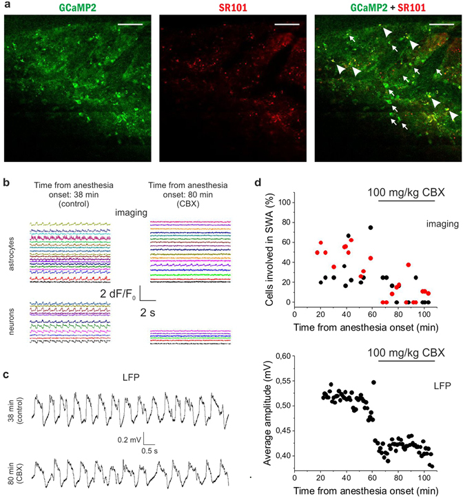
The gap junction blocker carbenoxolone (CBX) reduces the number of astrocytes and neurons taking part in the slow wave-associated network activity. (A) Expression of GCaMP2 (green), labeling of astrocytes with SR101 (red). Both neurons (some marked by arrows) and astrocytes (some marked by arrowheads) express GCaMP2. Scale bars: 100 µm (B) Left: 10-sec segments of ∆F/F0 fluorescent intensity traces of all identified astrocytes (n = 18) and neurons (n = 11) in the imaged area in the absence of CBX at 38 min following anesthesia. Right: 10-sec segments of ∆F/F0 fluorescent intensity traces of all identified astrocytes (n = 17) and neurons (n = 6) in the imaged area in the presence of CBX at 80 min following anesthesia. Different cells were imaged at 38 and 80 min. (C) Representative LFP recordings low-pass filtered at 20 Hz at different stages shown in B. (D) Top: Ratio of V1 neurons (black) and astrocytes (red) in the field of view showing repetitive Ca2+ transients under ketamine/xylazine anesthesia before and after i.p. administration of 100 mg/kg CBX. Individual markers represent data from each 60-sec imaging sessions. Bottom: Average amplitude of local field potential (LFP) positive deflections in each 1-minute segment of the LFP recording in the rat primary visual cortex V1 before and after i.p. administration of 100 mg/kg CBX. All data were recorded from the V1 area of a P44 rat (n = 24 imaging session, 60 s each).
