
Figure 7.
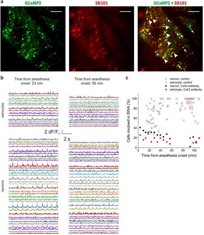
Specific blockade of the astrocytic gap junction subtype Cx43 inhibits synchronization in both astrocytes and neurons in vivo. (A) Expression of GCaMP2 (green), labeling of astrocytes with SR101 (red). Both neurons (some marked by arrows) and astrocytes (some marked by arrowheads) express GCaMP2. Scale bars: 100 µm (B) Left: 10-sec segments of ∆F/F0 fluorescent intensity traces of all identified astrocytes (n = 15) and neurons (n = 34) in the imaged area at 23 min following anesthesia. Right: 10-sec segments of ∆F/F0 fluorescent intensity traces of all identified astrocytes (n = 15) and neurons (n = 34) in the imaged area at 56 min following anesthesia. The same cells were imaged at 23 and 56 min. Cx43 antibody was applied 24 hours before the experiment. (C) The ratio of astrocytes (red) and neurons (black) showing repetitive, UP state-related Ca2+ transients in percentage of all astrocytes or neurons, respectively, in the field of views in the same P43 rat in the presence of Cx43 antibody (solid markers).Individual markers represent data from each 300-sec imaging sessions. Empty markers represent the ratio of astrocytes (red) and neurons (black) showing repetitive, UP state-related Ca2+ transients in the absence of Cx43 antibody. Note that large-scale synchronization is inhibited by the Cx43 antibody in both neurons and astrocytes. All data were recorded from the V1 area of a P43 rat (n = 15 imaging session, 300 s each).