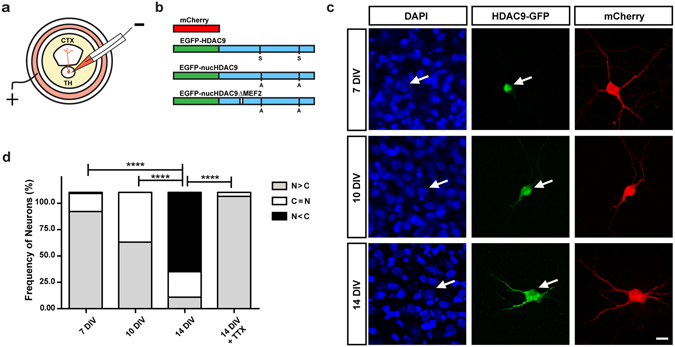
Figure 2.

Subcellular localization of HDAC9-EGFP fusion protein in TC organotypic co-culture preparations. (a) Experimental set-up of the co-culture preparation and electroporation into thalamic cells. (b) Schematic representation of the plasmids used in the present study (see the methods). (c) Representative thalamic neurons at 7, 10 and 14 DIV. HDAC9-EGFP distribution in the cells can be compared to the cytoplasmic protein mCherry and the nuclear staining of DAPI. Arrows refer to the position of the same cell at a given time point. Scale bar 10 μm. (d) Quantification of subcellular localization of HDAC9 was expressed as the ratio of transfected cells to illustrate HDAC9-EGFP signal concentrated predominantly in the nucleus (N > C), equally in the nucleus and the cytoplasm (N = C) or predominantly in the cytoplasm (N < C). Statistical analysis by overall Chi-square (Chi = 158, with 6 degrees of freedom, p < 0.000001) followed by pairwise Chi-square with Bonferroni correction. ****p < 0.0001.
