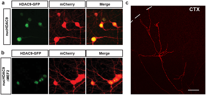
Figure 3.

Subcellular localization of HDAC9 mutants in thalamic cells and TC axon branching in vitro. Both of nucHDAC9-EGFP (a) and nucHDAC9ΔMEF2-EGFP (b) were distributed in the nuclei of thalamic cells at 14 DIV, while co-expressed mCherry was distributed in cell bodies and dendrites. Green: EGFP signal from the fusion protein. Red: mCherry. Scale bar, 5 μm. (c) Representative mCherry-labeled axon in the cortical explant, which elongated from the transfected thalamic cell. Interrupted line indicates the pial surface of the cortical explant. Scale bar 100 μm.
