Figure 6.

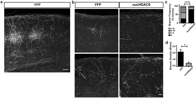
HDAC9 nuclear export enhances axon branching in vivo. (a) EYFP-labeled TC axonal projection in the P7 SSp cortex of an animal whose thalamus was electroporated with EYFP plasmid. Coronal section of approximately 60 μm thickness. Scale bar 100 μm. (b) Representative TC axon fragments from control (EYFP-electroporated) and nucHDAC9/mCherry co-electroporated neurons. Coronal sections of 20 μm thickness. Scale bar 50 μm. Statistical analysis by Chi-square (Chi = 22.3, with 3 degrees of freedom), p < 0.0001. (c) Distribution of axon segments according to their branch number. (d) Average branch density. Statistical analysis by Student t-test. *p < 0.05.
