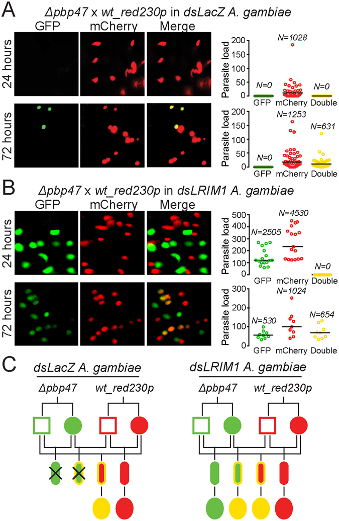
Figure 5.

A. gambiae co-infections with Δpbp47 and wt_red 230p parasite lines. Mosquitoes injected with dsLacZ (A) or dsLRIM1 (B) were fed on mice co-infected with GFP-expressing Δpbp47 and mCherry-expressing wt_red 230p parasites. Parasites in the mosquito midguts were enumerated at 24 and 72 hours post blood feeding. The left composite panel consists of representative fluorescence microscopy images (GFP, mCherry and combination of the two channels). The graphs on the right show the load of GFP-positive, mCherry-positive and GFP/mCherry double-positive parasites per midgut at each time point. Horizontal black lines indicate the median parasite load. Data from three independent biological replicates are pooled; N is the total number of parasites counted. (C) Genograms summarizing the results of A and B in dsLacZ (left) and dsLRIM1 (right) injected mosquitoes. Squares and circles correspond to male and female gametocytes, respectively; small ellipses correspond to ookinetes; large ellipses correspond to early stage oocysts. The outline color indicates the genotype: green is Δpbp47 homozygotes, red is wt_red 230p homozygotes and yellow is Δpbp47/wt_red 230p heterozygotes. The fill-in color indicates the phenotype: green is GFP-positive, red is mCherry-positive and yellow is GFP/mCherry double-positive parasites. Crossed parasites are killed and not detected.
