Figure 3.

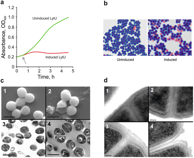
Overexpression of LytU. (a) The growth of S. aureus Newman strain containing an expression plasmid producing LytU. An immediate arrest of growth upon induction as compared to the uninduced culture was seen. These cultures were sampled for microscopic examinations. (b) Light microscope images of Gram-stained samples from a repeat for the one shown in panel (a) at the end time of 2 h 35 min when cultures were sampled for EM. A significant increase in a number of dead cells is seen in the induced culture. (c) Scanning EM and transmission EM study of the uninduced (1 and 3, respectively) and induced (2 and 4, respectively) samples. Scale bars, 1 µm. Images of the uninduced sample did not reveal any differences from normal growth thus excluding any defects caused by the presence of the expression vector or the uninduced insert. In scanning EM images, dead empty cells and cell debris are seen, explaining the stickiness of the sample and in accordance with Gram-staining results shown in panel (b). Multiple cell division sites are seen on the surface of the cells and transmission EM pictures clearly showed multiple daughter cells (up to about three divisions in maximum) within one mother cell. (d) Close-ups of the site of daughter cell separation. Scale bars, 50 nm. Image 1, uninduced cell are showing normal early-stage septa (plasma membranes formed) at the onset of CW synthesis. These kinds of divisions were very hard to find in the induced sample. Image 2, the later stage of a normal (uninduced) cell where the plasma membranes of daughter cells have started to separate and CW PG is accumulating in between. Normal progression inwards curving and restructuring of the CW surface that forms a surface groove is seen. Image 3, a cell where LytU has been induced. The CW between daughter cells is well formed, but no separation or reformation of mother cell’s CW is still seen. Image 4, the eventually broken CW of a LytU overexpressing cell in which the PG layer has not been restructured.
