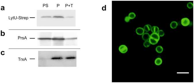
Figure 4.

Determination of orientation and localisation of LytU in the plasma membrane. (a, b, c) Trypsin treatment of protoplast surface proteins showing that LytU behaves like a protein outside on the plasma membrane surface. The three vertical immunoblotting lanes in the assay correspond to: P, protoplasts; PS, protoplast supernatant; P + T, protoplasts treated with 1 mg ml-1 trypsin. As the level of production from the used expression vector is controlled by the amount of xylose inducer, 0.02% xylose was used to get very low amounts of LytU, resembling the natural conditions. The controls are shown in Supplementary Fig. S1. (d) Confocal microscopy image of S. aureus cells showing fluorescence of the N-terminal GFP-fusion to LytU protein. Scale bar, 2 μm. Clear localization of the fusion protein in the plasma membrane is seen as well as some possible accumulation at cell division sites (see text and Methods for details).
