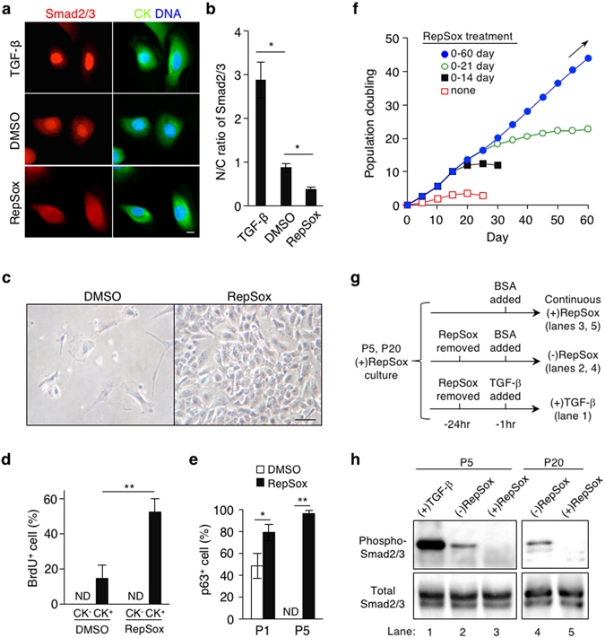
Figure 1.

Inhibition of TGF-β signaling enables long-term expansion of p63+ mouse primary epidermal progenitor cells in vitro. (a) Subcellular localization of Smad2/3 in newborn mouse primary epidermal keratinocytes grown in CnT-PR media. The cells were stimulated with 1 ng/ml TGF-β2, 0.1% DMSO (as a control) or 1 μM RepSox for 1 hr. CK, pan-cytokeratin; DNA, nuclear counterstaining with Hoechst 33342; Bar = 10 μm. (b) Nuclear-to-cytoplasmic (N/C) ratios of Smad2/3 as determined by fluorescence intensity. Data shown are mean ± s.e.m. (n = 6). *P < 0.05. (c) Representative images of primary cells of newborn mouse epidermis at day 14 of culture in the presence or absence of 1 μM RepSox. Bar = 25 μm. (d) A short-pulse labeling with BrdU in primary culture of newborn mouse epidermal cells in the presence or absence of 1 μM RepSox. Data shown are % per field, expressed as mean ± s.e.m. (n = 4). ND, not detected; **P < 0.01. (e) Percentages of p63+ cells at passage 1 and 5 (P1 and P5, respectively) grown in the presence (solid) or absence (open) of 1 μM RepSox. Data shown are % per field, expressed as mean ± s.e.m. (n = 3 and 4 for P1 and P5, respectively). ND, not detected; *P < 0.05; **P < 0.01. (f) Population doubling of newborn mouse-derived, RepSox-expanded P2 epidermal cells grown in CnT-PR media in the presence of 1 μM RepSox for 0, 14, 21, and 60 days. Data shown are representative of three independent experiments with similar results. Arrow indicates continuous cell growth. (g,h) Removal of RepSox associates with increased Smad2/3 phosphorylation. (g) Experimental design. Newborn mouse-derived, RepSox-expanded P5 and P20 epidermal keratinocytes were further grown in continuous presence (upper) or absence (middle and lower) of 1 μM RepSox for 24 hrs. Culture of P5 cells stimulated with 1 ng/ml TGF-β for 1 hr prior to lysis (lower) was used as a positive control. (h) Expression of total and phosphorylated Smad2/3 as determined by Western blot. Lane numbers correspond to those in (g). The full-length blots are included in Supplementary Fig. S11.
