Figure 2.

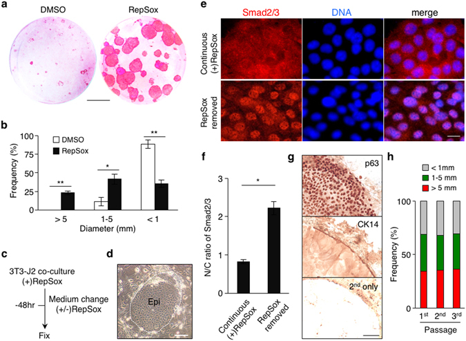
Inhibition of TGF-β signaling supports self-renewal of mouse primary epidermal progenitor cells in serial 3T3-J2 co-culture. (a) Rhodamine B staining of newborn mouse-derived, RepSox-expanded P1 epidermal cells grown in 3T3-J2 co-culture in the presence or absence of 1 μM RepSox for 14 days. Bar = 10 mm. (b) Distribution of epidermal clone sizes at day 14. Data shown are mean ± s.e.m. (n = 3). *P < 0.05; **P < 0.01. (c–f) Removal of RepSox associates with nuclear translocation of Smad2/3 in newborn mouse epidermal cells in 3T3-J2 co-culture. (c) Experimental scheme. Mouse primary epidermal keratinocytes were grown in 3T3-J2 co-culture in the presence of 1 μM RepSox. Subsequently, the cultures were maintained in the presence or absence of 1 μM RepSox for 48 hrs before fixation for immunohistochemistry. (d) A representative image of mouse primary keratinocyte clones in 3T3-J2 co-culture. Note that epidermal cell clones show a cobblestone appearance with a distinct clone border (dotted line). Epi, epidermal cell clone. Bar = 50 μm. (e) Representative immunofluorescence images of mouse epidermal cells without (upper) or with (lower) removal of RepSox, stained with anti-Smad2/3 antibodies (left panels) and counterstained with Hoechst 33342 (middle panels). Right panels show merged images. Bar = 10 μm. (f) Nuclear-to-cytoplasmic (N/C) ratios of Smad2/3 as determined by fluorescence intensity. Data shown are mean ± s.e.m. (n = 10). *P < 0.001. (g) Immunohistochemistry of representative epidermal cell clones grown in 3T3-J2 co-culture for 14 days in the presence of 1 μM RepSox, stained with anti-p63 (upper) and anti-CK14 (middle) antibodies. The lower panel represents a control clone stained with the secondary antibodies alone. Data shown are peripheral regions of three different epidermal clones with similar diameter. Bar = 100 μm. (h) Distribution of epidermal clone sizes in serial co-culture with 3T3-J2 cells in the presence of 1 μM RepSox.
