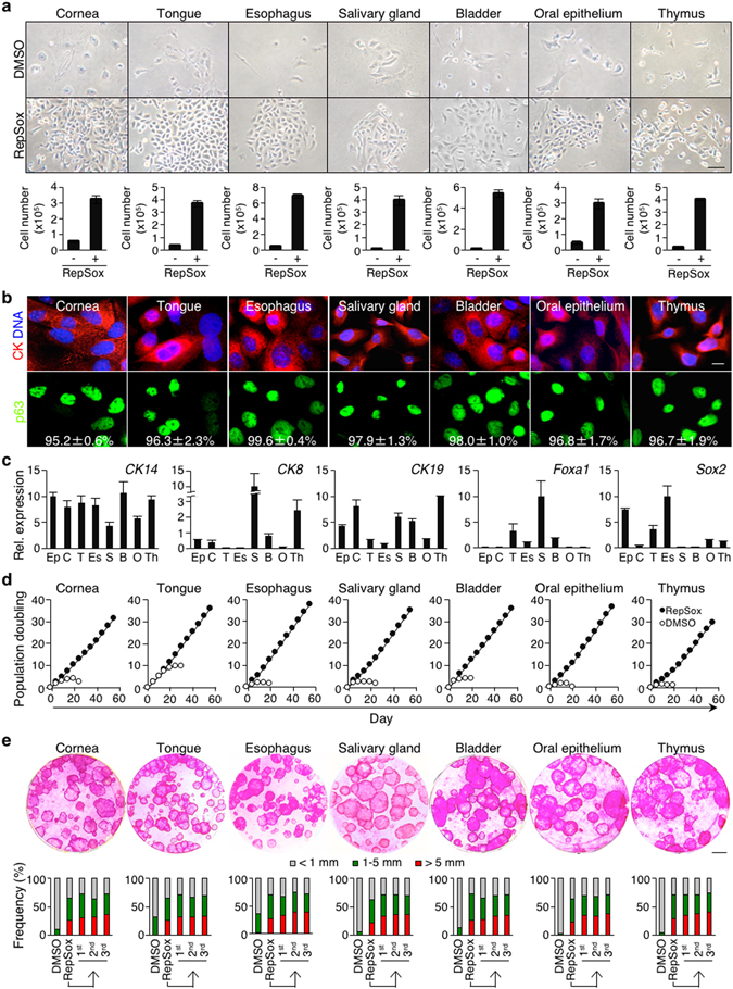
Figure 5.

Inhibition of TGF-β signaling enables long-term expansion of p63+ epithelial progenitor cells of diverse mouse epithelia. (a) Representative images of primary cells of various newborn mouse epithelia, grown in the presence (lower) or absence (upper) of 1 μM RepSox for 6 days. Bar = 50 μm. Bar graphs below indicate numbers of primary cells at day 7 of culture. CnT-PR-expanded CK+ mouse primary epithelial cells (2 × 10⁴) were seeded. Data shown are mean ± s.e.m. (n = 3). (b) Representative immunofluorescence images of newborn mouse-derived, RepSox-expanded P2 epithelial cells stained with anti-p63 and anti-pan-CK antibodies and counterstained with Hoechst 33342 (DNA). Numbers shown in lower panels represent percentages of p63+ cells per field, expressed as mean ± s.e.m. (n ≥ 4). Bar = 10 μm. (c) Quantitative RT-PCR analysis of epithelial progenitor cell-associated genes using newborn mouse-derived, RepSox-expanded P9 (thymus and salivary gland), P10 (epidermis), and P11 (cornea, oral epithelium, tongue, esophagus and bladder) cells. For each gene, the highest expression was set to 10. Data shown are normalized to the housekeeping gene Rps18 and expressed as mean ± s.e.m. (n = 3). Ep, epidermis; C, cornea; T, tongue; Es, esophagus; S, salivary gland; B, bladder; O, oral epithelium; and Th, thymus. (d) Population doubling of newborn mouse-derived, RepSox-expanded P2 epithelial cells grown in CnT-PR media in the presence (solid) or absence (open) of 1 μM RepSox. (e) 3T3-J2 co-culture. Representative images of Rhodamine B staining of epithelial clones grown for 14 days in the presence of 1 μM RepSox. Newborn mouse-derived, RepSox-expanded P1 epithelial cells were used. Bar = 5 mm. Data shown below indicate the distribution of epithelial clone sizes during serial 3T3-J2 co-cultures. Epithelial cells harvested from RepSox-treated primary 3T3-J2 co-cultures (lane 2) were serially cultivated with two-week intervals with fresh 3T3-J2 cells in the presence of 1 μM RepSox (lanes 3–5). DMSO, primary cultures without RepSox (lane 1).
