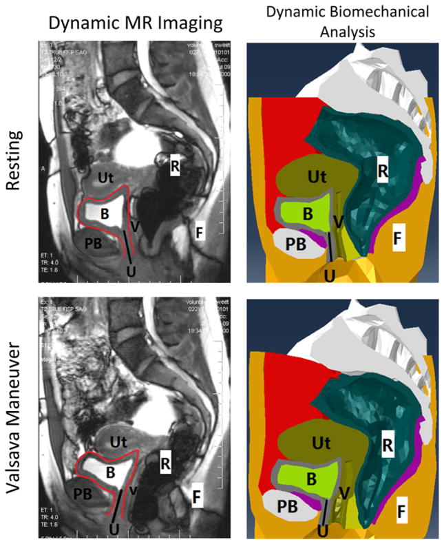
Figure 2.
Comparison (dynamic MR imaging vs. dynamic biomechanical analysis) of the pelvic structures of the female subject in the sagittal plane, at resting stage and at Valsalva stage. The black solid line in all pictures shows the location of urethra. The red curves in the dynamic MRI outline the bladder. (Abbreviations: Ut - Uterus, R - Rectum, B - Bladder, PB - Pubic Bone, V - Vagina, F - Fat, U - Urethra.)

