
Figure 2.
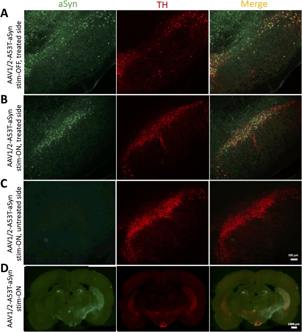
AAV1/2‐A53T‐aSyn injection into the SN leads to widespread expression of pathological A53T‐aSyn that projects into the striatum. (A,B) Representative immunofluorescence double stainings showing expression of A53T‐aSyn (green) and TH+ dopaminergic neurons (red) in the SN after 43 days of AAV injection in a (A) stim‐OFF rat of the AAV1/2‐A53T‐aSyn‐treated hemisphere and a (B) stim‐ON AAV1/2‐A53T‐aSyn‐treated rat revealing a widespread A53T‐aSyn expression in the SN of both rats and more surviving TH+ cells in the AAV1/2‐A53T‐aSyn stim‐ON brain. (C) No detectable aSyn aggregates in the SN of the untreated side of the AAV1/2‐A53T‐aSyn stim‐ON rat are visible. (D) Coronal whole‐brain slice of a AAV1/2‐A53T‐aSyn stim‐ON rat shows the deposition of aSyn (green) and nigrostriatal dopaminergic projections (red) in the dorsal striatum. AAV = adeno‐associated virus; aSyn = α‐synuclein; SN = substantia nigra; TH = tyrosine hydroxylase.