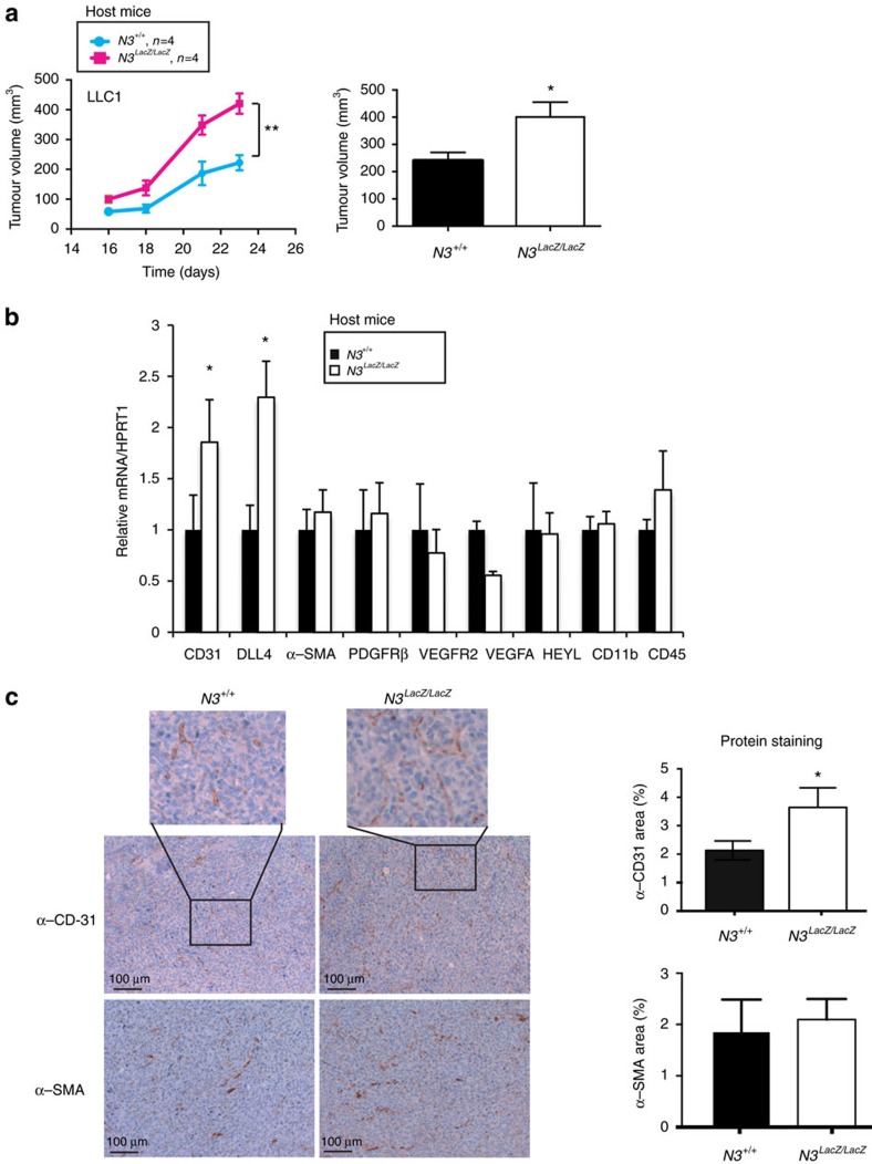
Figure 2. Notch3 limits tumour growth and vascularization in vivo.
(a) 5 × 10⁵ LLC1 cells were implanted into the left flank of wild-type C57Bl/6 mice (N3+/+, n=4) or of Notch3 LacZ homozygous Knock-in C57Bl/6 littermates (N3LacZ/LacZ, n=4). Tumour growth was monitored from day 16 until day 24 when mice were sacrificed. Two-way ANOVA was performed to assess time and genotype effect on tumour growth (Interaction: P=0,013; Time: P<0,0001; Genotype: P=0,0015). (b) mRNA was extracted from tumours dissected after 14 days of growth from wild-type C57Bl/6 mice (N3+/+, n=6) or Notch3 mutant mice N3LacZ/LacZ C57Bl/6 littermates (N3LacZ/LacZ, n=5). Quantitative RT–PCR was performed to measure CD31, DLL4, PDGFRβ, α-SMA, VEGFR2, VEGFA, CD11b and CD45 expression (means±s.d., unpaired t-test was applied). (c) Immunohistochemistry for CD31 and α-SMA was performed on tumours dissected from wild-type mice (N3+/+, n=15) or Notch3 mutant (Notch3LacZ/LacZ) mice littermates (n=9) on day 14. Images are representative of four different sections from each tumour. Quantification was done using ImageJ angiogenesis plug-in on four different images from each tumour (mean±s.e.m. for quantification, unpaired t-test).

