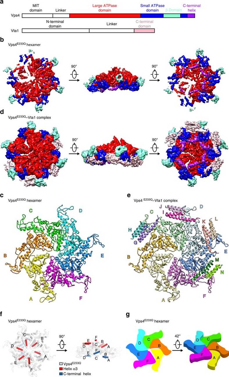
Figure 1. Overall structures of the ATP-bound Vps4E233Q hexamer and its complex with Vta1.
(a) Domain diagrams of the yeast Vps4 (upper) and Vta1 (lower). (b) Different views of the EM density map of the ATP-bound Vps4E233Q hexamer at 5.8 σ contour level. (c) The atomic model of the Vps4E233Q hexamer. Vps4E233Q subunits are colour coded. (d) Different views of the EM density map of the ATP-bound Vps4E233Q–Vta1 complex at the 3.9 σ contour level showing the positions of the Vta1. (e) The atomic model of the ATP-bound Vps4E233Q–ta1 complex. Vps4E233Q subunits and Vta1 subunits are colour coded. (f) Different views of the helix α3 and C-terminal helix in the Vps4E233Q hexamer to show the spiral arrangement of Vps4 subunits. (g) A schematic diagram showing the topology of the Vps4E233Q subunits in the Vps4E233Q hexamer. The colouring scheme is the same as in (c).

