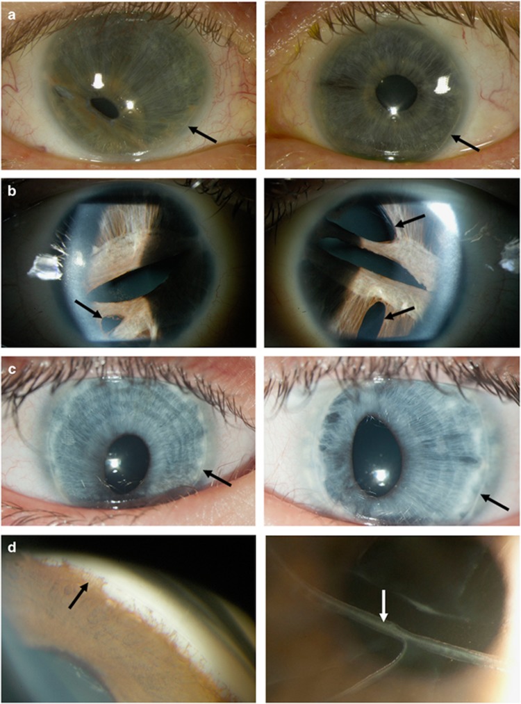
Figure 3.
Clinical photographs of individuals with ocular features of Axenfeld-Rieger Malformation. Photographs in (a–c) showing the right eye (left panel) and the left eye (right panel). (a) Slit lamp photos showing corectopia in the left panel, iris stromal hypoplasia in both eyes and posterior embryotoxon (black arrows) in both eyes (individual 9B). (b) Slit lamp photos showing corectopia, pseudopolycoria (black arrows) and iris stromal hypoplasia in both eyes (individual 18B). (c) Slit lamp photos showing corectopia and posterior embryotoxon (black arrows) in both eyes (individual 12). (d) Gonioscopy showing irido-corneal adhesions (black arrow, left panel) and photo showing the presence of breaks in the Descemet’s membrane (Haab’s striae, white arrow, right panel; individual 5A).

