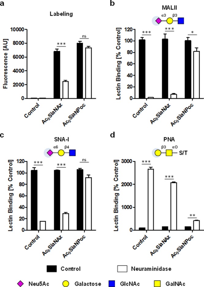
Figure 3.

Enzymatic removal of Az and Poc sialic acids from the cell surface of THP-1 cells. Cells incubated for 3 days with PBS, 100 μM Ac5SiaNAz, or 100 μM Ac5SiaNPoc were treated for 1 h with 250mU/mL Clostridium perfringens neuraminidase. Az and Poc sialoglycans were reacted to fluorescent biotin using CuAAC (a), α2,3-sialoglycans were detected with MALII lectin (b), α2,6-sialoglycans were detected with SNA-I lectin (c), and terminal β-galactose was detected with PNA lectin (d). Bar diagrams show mean fluorescence intensity or mean lectin binding normalized to control ± SEM of three independent experiments. MALII: Maackia amurensis lectin; PE: phycoerythrin, PNA: Peanut agglutinin lectin; SEM: standard error of the mean; SNA-I: Sambucus nigra lectin.
