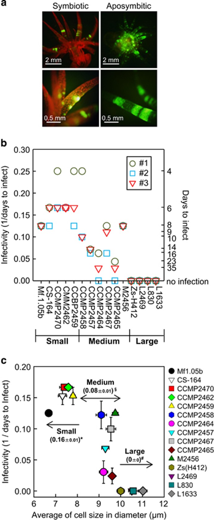
Figure 2.
Infection of different Symbiodinium strains into Aiptasia. (a) Fluorescence stereomicroscope images of Aiptasia anemones with (symbiont) and without (aposymbiont) symbiotic algae Symbiodinium. Red fluorescence is from the chlorophyll in Symbiodinium, while the autofluorescence of Aiptasia is green. (b) Days to infect (the number of days until infection was verified) and infectivity (1/days to infect) are shown on the right and left y axes, respectively. An Aiptasia polyp was said to be infected when >30 foci of Symbiodinium cells could be seen within a tentacle. Three independent experiments (#1, #2 and #3) were carried out in each Symbiodinium strain. (c) Relationship between Symbiodinium cell size and infectivity. Numbers in the panel show the average infectivity (1/days to infect)±s.e. of each size group. Different symbols (*, § and #) indicate significant difference (P<0.01) between groups. Error bars, ±s.e. (n=3).

