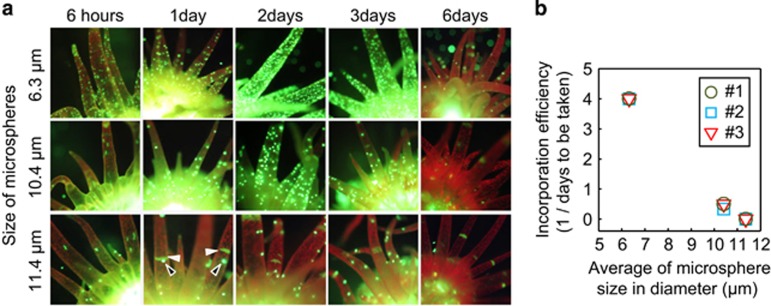
Figure 3.
Incorporation of microspheres of different sizes into Aiptasia. (a) Fluorescence stereomicroscopic images of Aiptasia anemones after introducing yellow-green fluorescent microspheres of different sizes (6.3±0.18, 10.4±0.25, and 11.4±0.3 μm in diameter). Microspheres of each size were separately fed into the mouths of anemones and their presence in tentacles was monitored for 6 days. White and black arrow heads indicate autofluorescence of Aiptasia and microspheres, respectively. (b) Relationship between incorporation efficiency (1/the number of days until incorporation was verified) and average microsphere size. An Aiptasia polyp was said to have incorporated microspheres when >30 foci could be seen within a tentacle. Three independent experiments (#1, #2 and #3) were carried out in each size.

