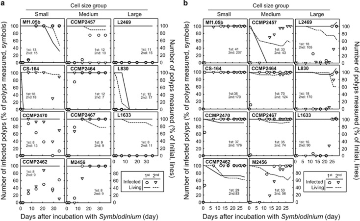
Figure 5.
Infection of different Symbiodinium strains into corals. Aposymbiotic juvenile polyps of (a) A. tenuis and (b) C. serailia were incubated with different Symbiodinium strains and monitored for 35 and 30 days, respectively. Number of infected polyps is shown on the left y axes (percentage of total polyps measured, symbols). Number of polyps measured at each time point (percentage of initial, lines) is shown on the right y axes. Only healthy looking polyps were included in the measurements at each time point. A coral polyp was said to be infected when >30 foci of Symbiodinium cells could be seen within a tentacle. Each experiment was carried out twice using two set of polyps in different containers (microplates) and each result is shown separately. First experiment, closed symbols and solids line. Second experiment, open symbols, dashed line. The initial number of polyps used for the first and second experiment is shown in each panel.

