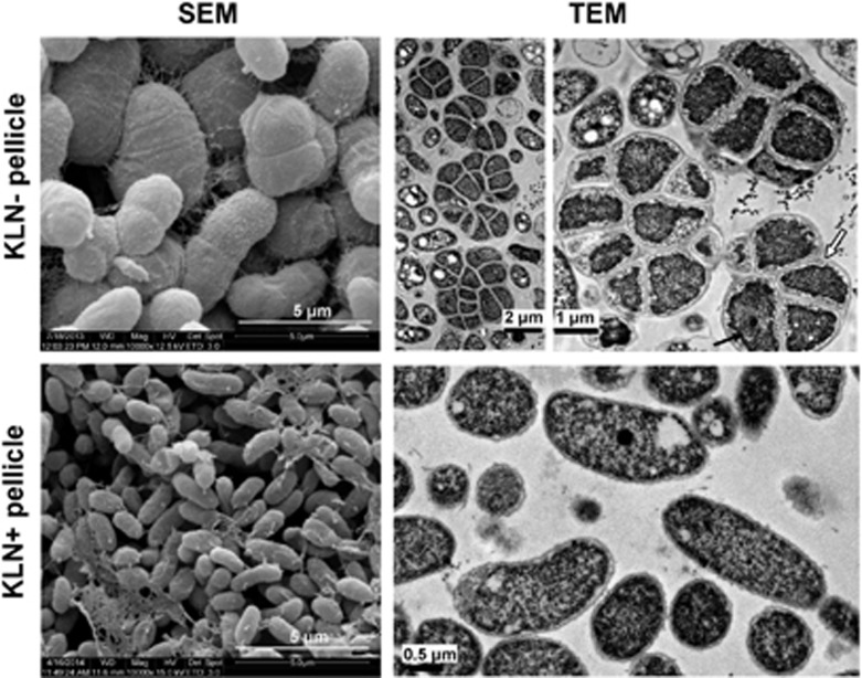
Figure 6.
SEM and TEM images of Azospirillum brasilense Sp7 pellicles formed in KLN− and KLN+ media. The A1501 cyst-like large ‘cells’ (EPS-encased bacterial aggregate) were found in KLN− pellicles, but not from KLN+ pellicles. Black arrow in TEM picture of KLN− pellicle indicates the bacteria cell inside the EPS sac, and white arrow indicates the edge of EPS sac. Two TEM images of A1501 cyst-like structures from Sp7 KLN− pellicle were shown in the upper panel.

