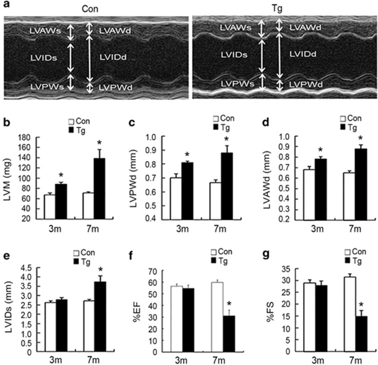
Figure 2.
Cardiac dysfunction in miR-199a transgenic mice. (a) Representative M-mode images of a miR-199a Tg mouse (right) and the wild-type littermate control (left). (b) Quantification of LV mass. (c and d) Measurements of the LV wall thickness in diastole (LVPWd and LVAWd). (e) Measurements of the LV internal diameters in systole (LVIDs). (f and g) Quantification of ejection fraction (EF) and fractional shortening (FS) in the hearts from miR-199a Tg mice and control mice at 3 and 7 months. Mean values±SEM were determined by echocardiography. 3-Month-old control mice: n=5; 3-month-old Tg mice: n=6; 7-month-old control mice: n=7; 7-month-old Tg mice: n=8; *P<0.05

