Figure 4.
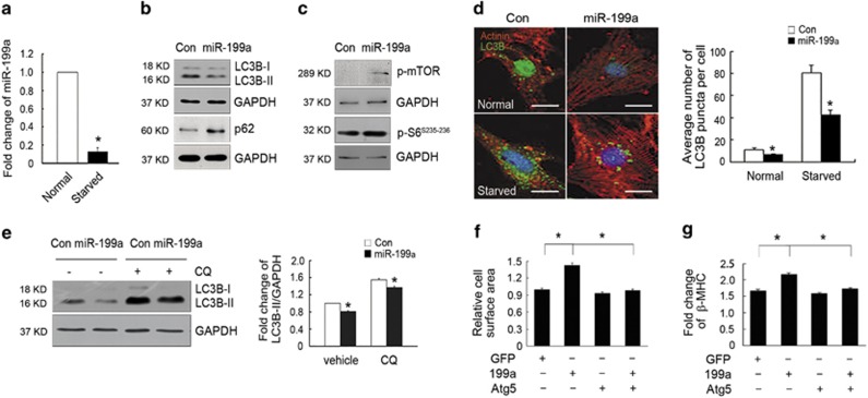
miR-199a inhibit cardiomyocyte autophagy in a cell-autonomous manner. (a) Real-time PCR analyses of miR-199a expression under normal or serum/glucose-deprivation (starved) conditions. (b and c) Western blotting analyses of LC3B-I, LC3B-II, p62 and mTOR/S6 protein levels in cardiomyocytes infected with adenoviruses expressing control or miR-199a miRNAs. (d) Representative images and quantification of GFP-LC3B puncta (green) in control and miR-199a-overexpressing cardiomyocytes under normal or starved conditions (n=30 cells/per condition). Scale bars represent 5 μm. (e) Representative western blotting and quantification showing reduced LC3B-II levels in miR-199a overexpression cardiomyocytes treated with or without CQ. (f) Atg5 overexpression rescued the miR-199a-induced hypertrophic growth of cardiomyocytes. Fold change in mean cell surface area of α-actinin-immunostained cardiomyocytes infected with adenoviruses (n=160). *P<0.01. (g) Real-time PCR showing that Atg5 overexpression rescued β-MHC upregulation induced by miR-199a overexpression (n=3). *P<0.05. GAPDH, glyceraldehyde 3-phosphate dehydrogenase

