Figure 1.
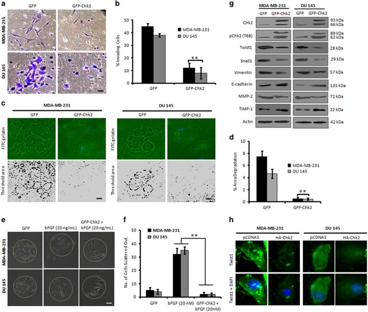
Chk2 expression negatively regulates invasion of p53-deficient cancer cells. (a) MDA-MB-231 and DU 145 cells were transiently transfected with GFP and GFP-Chk2 plasmid construct for 48 h and checked for the invasion of cells through matrigel invasion assay. Invading cells were observed and photographed under an inverted microscope at × 20 magnification. Scale bar: 20 μm. (b) Bar graph showing results from quantification of invading cells (n=3, error bars indicate±S.D.).**P<0.01. (c) Cells were transfected with GFP and GFP-Chk2 construct and assessed for their invadopodia formation and matrix degradation ability over FITC-gelatin matrix coverslips. Blue parts show the nucleus stained with DAPI mounting media. Images were taken at × 20 magnification, and the threshold area of degradation was determined with the help of Image J software. Scale bar: 20 μm. (d) Bar graphs showing the percent area of degradation quantified through Image J analysis (n=3, error bars indicate±S.D.).**P<0.01. (e) MDA-MB-231 and DU 145 cells were allowed to form the distinct colonies for 5 days, stimulated with bFGF (20 ng/ml) and then transfected with either GFP or GFP-Chk2 for 48 h. Cells were then observed for their scattering out of the colonies and photographed under × 20 magnification. Scale bar: 20 μm. (f) A total of five random colonies from each field were analyzed and counted manually under the microscope for the number of scattered cells and was adjusted to the total cells in particular colony. Bar graphs showing quantification of the average number of cells scattered out of the colonies (n=3, error bars indicate±S.D.).**P<0.01. (g) Cells were transfected with GFP or GFP-Chk2 for 48 h; whole-cell lysates were prepared, and an equal amount of protein (20 μg) was subjected to western blot analysis for the expression of mesenchymal markers like; Twist1, Snail1, vimentin, MMP-2 and epithelial markers such as E-cadherin and TIMP-1 (n=3). (h) Immunofluorescence staining of Twist1 in MDA-MB-231 and DU 145 cells after transiently transfecting the cells with pCDNA3 and HA-Chk2 for 48 h (original magnification × 20)

