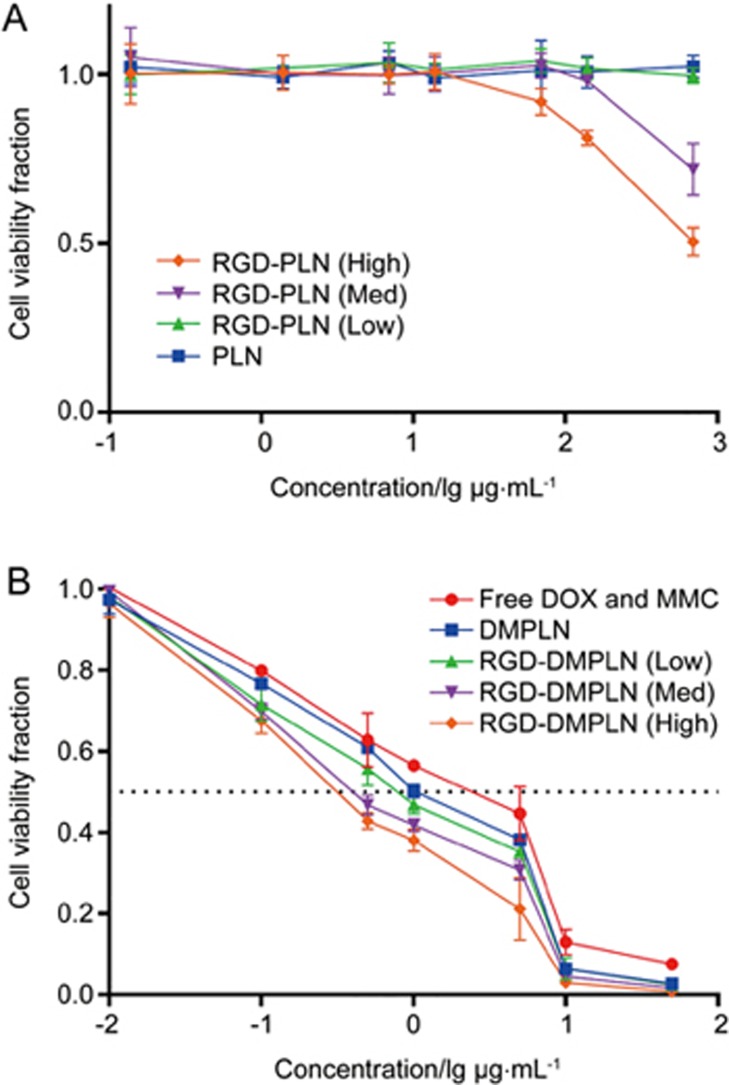
Figure 2.
In vitro cytotoxicity of particles in MDA-MB-231-luc-D3H2LN cells. Cells were exposed to seven concentrations of (A) blank NPs (PLN: 0.139–695 μg/mL) or (B) free and nanoparticle DOX and MMC formulations (DOX: 0.01–50 μg/mL) for 1 h. In both (A) and (B) cells were washed and allowed to proliferate for 24 h before evaluated by ATP bioluminescence assay. At [DOX] of 50 μg/mL in NP formulation, the corresponding PLN is 695 μg/mL. All formulations of DOX and MMC were given at the DOX/MMC ratio of 1: 0.7. Data are presented as mean±SD. n=3 for each formulation.

