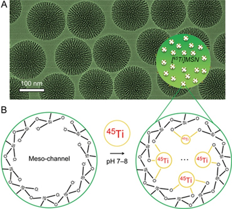
Figure 2.
Synthesis and characterization of MSN. (A) A transmission electron microscopy (TEM) image of ∼150 nm sized MSN particles. Surface area: 581.5 m2/g. Pore volume: 1.36 cm3/g. Average pore size: 4–5 nm. (B) A schematic illustration showing the labeling of 45Ti to the deprotonated silanol groups (-Si-O-) from the outer surface and inner meso-channels of MSN. Please note the real coordination chemistry of 45Ti4+ with the silanol groups could be much more complex than the scheme shown in (B).

