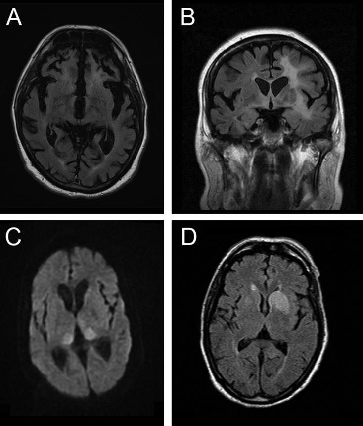
Figure 3.

MR scan of brain in four patients presenting as mimics of prion disease. (A) C9orf72 mutation showing severe generalised atrophy with no parenchymal abnormal signal. Severe atrophy does occur late in some cases of Creutzfeldt-Jakob disease (CJD), but usually diffusion-weighted sequences reveal restricted diffusion. (B) B-cell lymphoma confined at the time of presentation to the brain. Note the extensive white-matter signal change with the normal cortex. Occasionally, the leukoencephalopathic form of CJD has white-matter signal alteration, but this is associated with grey matter destruction. (C) N-methyl-D-aspartate antibody encephalitis with MR imaging showing the pulvinar sign. This sign develops in most cases of variant CJD and has rarely been described with other pathologies. (D) Voltage-gated potassium channel (Casp-1) antibody encephalitis showing unilateral basal ganglia high signal on diffusion-weighted imaging but not on the apparent diffusion coefficient map (scan not shown). Note that the swelling of the caudate is a feature that does not occur in CJD.
